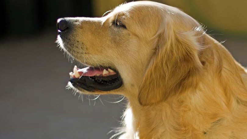
Hip Dysplasia in Dogs
Hip dysplasia is one of the most common problems afflicting large and giant breeds of dogs, including Germans shepherds, labradors and golden retrievers, and Rottweilers. Males are at a greater risk for developing the disease than females, possibly because of their larger size and more rapid growth. The primary cause of hip dysplasia is genetic; dogs with parents and other relatives with dysplasia are more likely to develop the condition themselves. Unfortunately, the trait is passed from generation to generation in a complicated manner, so determining exactly which animals are at risk is not simple. Puppies that are fed a diet extremely rich in calories, protein, and calcium also have an increased likelihood of developing hip dysplasia.
The condition arises when one or both of a dog’s hips fail to develop normally. The hip is a “ball and socket” type of joint composed of the femoral head, a round structure at the top of the femur, and the acetabulum, which is a cup-shaped depression in the pelvis. A normal acetabulum surrounds about two-thirds of the femoral head and grips it securely. Cartilage and fluid within the joint allow to the femoral head to rotate smoothly and easily within the acetabulum. In cases of hip dysplasia, the acetabulum is flatter than it should be, which allows the femoral head to rattle around within the joint. This excess motion leads to inflammation and pain. With time the body tries to reduce the abnormal motion by depositing bone around the joint, a painful condition known as osteoarthritis.
Symptoms
A dog with hip dysplasia will typically favor the hind leg that is most severely affected. However, because both hips are often involved, an obvious limp is not always evident. Dogs may also have difficulty rising, jumping, and climbing stairs; can exhibit a rolling motion to their hind end when walking, bunny hoping when running or climbing stairs; be generally less active than normal; and lose muscle mass in their hind end. Young dogs that have not yet developed a lot of arthritis may not show many symptoms, even if their hips are severely abnormal.
Diagnosis
To differentiate hip dysplasia from other problems that can cause hind end lameness or weakness, a veterinarian will need to know what clinical signs an owner is seeing at home and to perform a thorough physical exam. The vet will assess the dog’s stride by watching it walk and trot from several different angles. The doctor will also flex, extend, and rotate the dog’s hips. In many cases, dogs with hip dysplasia do not exhibit pain when their upper hind leg is pressed towards their abdomen, but if the leg is pulled backward or rotated away from the body, they become obviously uncomfortable. Another method of determining whether hip dysplasia is likely is to evaluate a dog for an Ortolani sign. During this procedure, a dog must lie on its back with both hind legs pointing straight up. With hip dysplasia, the femoral heads will move away from their acetabula. As the veterinarian pushes the dog’s lower leg to the side, the femur can be felt and sometimes even heard popping back into its socket.
X-rays, which are also called radiographs, are necessary to definitively diagnose hip dysplasia, to assess its severity, and to determine if osteoarthritis is present. Many dogs will need to be sedated before being radiographed because the positions necessary to evaluate the hips can be painful. Severe cases of early hip dysplasia and osteoarthritis can be diagnosed with regular x-rays. However, milder cases may not be evident without specialized techniques.
PennHIP evaluations can be performed by veterinarians who have undergone training in the procedure. Dogs are radiographed with their femoral heads pushed into and pulled out of their acetabula. If a dog’s hips are very loose, dysplasia is more likely than if little change occurs with these manipulations. Dogs can be evaluated using the PennHIP technique after 16 weeks of age. The Orthopedic Foundation of America (OFA) rates dog hips as normal (excellent, good or fair), borderline, or dysplastic (mild, moderate or severe) based on x-rays, but dogs must be at least 24 months old to participate. Owners must also remember that normal x-rays, including PennHIP and OFA evaluations, do not completely eliminate the chance that dysplasia is present.
Treatment
If a dog is diagnosed with hip dysplasia when it is between 8 and 18 months of age, and it has not yet developed significant arthritis, and its acetabulum is not too flat, a triple pelvic osteotomy (TPO) surgery can reduce the amount of arthritis that would otherwise develop. With this technique, a veterinary surgeon cuts the pelvis in three places and realigns the acetabulum so that the femoral head is more securely held in place. Other surgical techniques are also available for young dogs, but they are not routinely performed.
If a dog has developed significant osteoarthritis and can’t be kept comfortable with pain-relievers and anti-inflammatory medications, either a femoral head and neck ostectomy (FHO) or a total hip replacement should be considered. During an FHO, the veterinarian will completely remove the femoral head and its supporting bone. With time, a false joint develops that allows the dog to walk, run, and play almost normally and without any pain. This surgery works best for dogs that are fairly active and weigh less than 50 pounds. Larger dogs do better when the hip joint is completely removed and artificial replacement parts are inserted into the femur and pelvis.
If surgery is not an option for a dog with hip dysplasia, medications that reduce pain and inflammation and supplements that protect cartilage can greatly improve a pet’s comfort and activity level. Acupuncture, cold lasers, and physical therapy can also be very useful. If a dog is too heavy, weight loss is one of the most effective methods of improving its quality of life. Moderate exercise, especially swimming and regular, slow walks, can help keep the structures that support a dog’s hips strong and functioning well.
To reduce the prevalence of hip dysplasia, many breeders have their animals tested through OFA or PennHIP. Puppies that are born to parents with good hips are less likely to develop dysplasia themselves. Feeding young, at risk dogs an appropriate amount of a food specifically designed for large breeds can also promote healthy hip development.
Prognosis
The various surgical techniques that are commonly used to treat hip dysplasia are very effective at keeping dogs comfortable and active as they age, but surgery may not be the right option for every owner or every dog. Nonsurgical therapy for hip dysplasia and its resulting osteoarthritis is a long-term endeavor. As a dog’s condition progresses over time, satisfactory pain control can become more difficult to accomplish.
Article By:
Jennifer Coates, DVM graduated with honors from the Virginia-Maryland Regional College of Veterinary Medicine in 1999. In the years since, she has practiced veterinary medicine in Virginia, Wyoming, and Colorado and is the author of several short stories and books, including the Dictionary of Veterinary Terms, Vet-Speak Deciphered for the Non-Veterinarian. Dr. Coates lives in Fort Collins, Colorado with her husband, daughter, and a menagerie of pets.
Having discovered a fondness for insects while pursuing her degree in Biology, Randi Jones was quite bugged to know that people usually dismissed these little creatures as “creepy-crawlies”.